Rev Bras Oftalmol.2026;85:e0075
Trabeculitis during herpetic uveitis: anterior segment optical coherence tomography findings

DOI: 10.37039/1982.8551.20260075
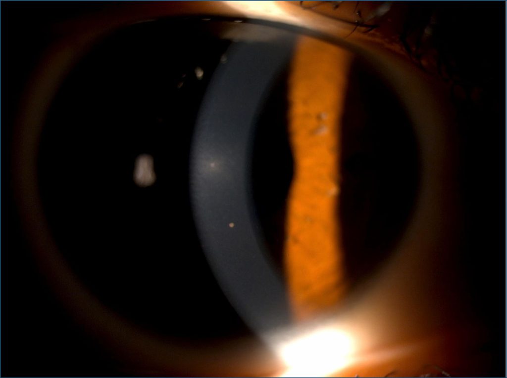
A 34-year-old man presented with pain and blurring vision in the right eye for 24 hours. The best-corrected visual acuity was 9/10 in the right eye. Slit-lamp examination of the right eye revealed conjunctival injection, three white granulomatous corneal precipitates (), one of them behind a stromal infiltrated area. A white tyndall effect in the anterior chamber at one cross and ocular hypertension at 50 mmHg were noticed. Gonioscopy shows a 360° open angle with a flat iris insertion root. Anterior segment optical coherence tomography (AS-OCT) revealed globular corneal precipitates appearing as protrusions attached to the endothelium (), floating cells in the anterior chamber and hyper-reflectivity of the trabecular meshwork due to the presence of cellular infiltration (). The diagnosis of likely herpetic uveitis was made. The patient received valacyclovir 3 g per day with hypotonizing treatment. The intraocular pressure of the right eye was 17 mmHg after 2 days. Topical corticosteroids were added to the treatment with full improvement of symptoms and signs of herpetic uveitis.
Granulomatous corneal precipitates and stromal infiltrate are typical signs of herpetic uveitis, especially when associated to intraocular hypertension. In this case, we report hyper-reflectivity of the trabecular meshwork as tomographic sign of trabeculitis associated to herpetic uveitis. Trabeculitis would be the cause of ocular hypertension. This sign is rarely reported.()
[…]