Rev Bras Oftalmol.2026;85:e0007
Herpes zoster keratitis
DOI: 10.37039/1982.8551.20260007
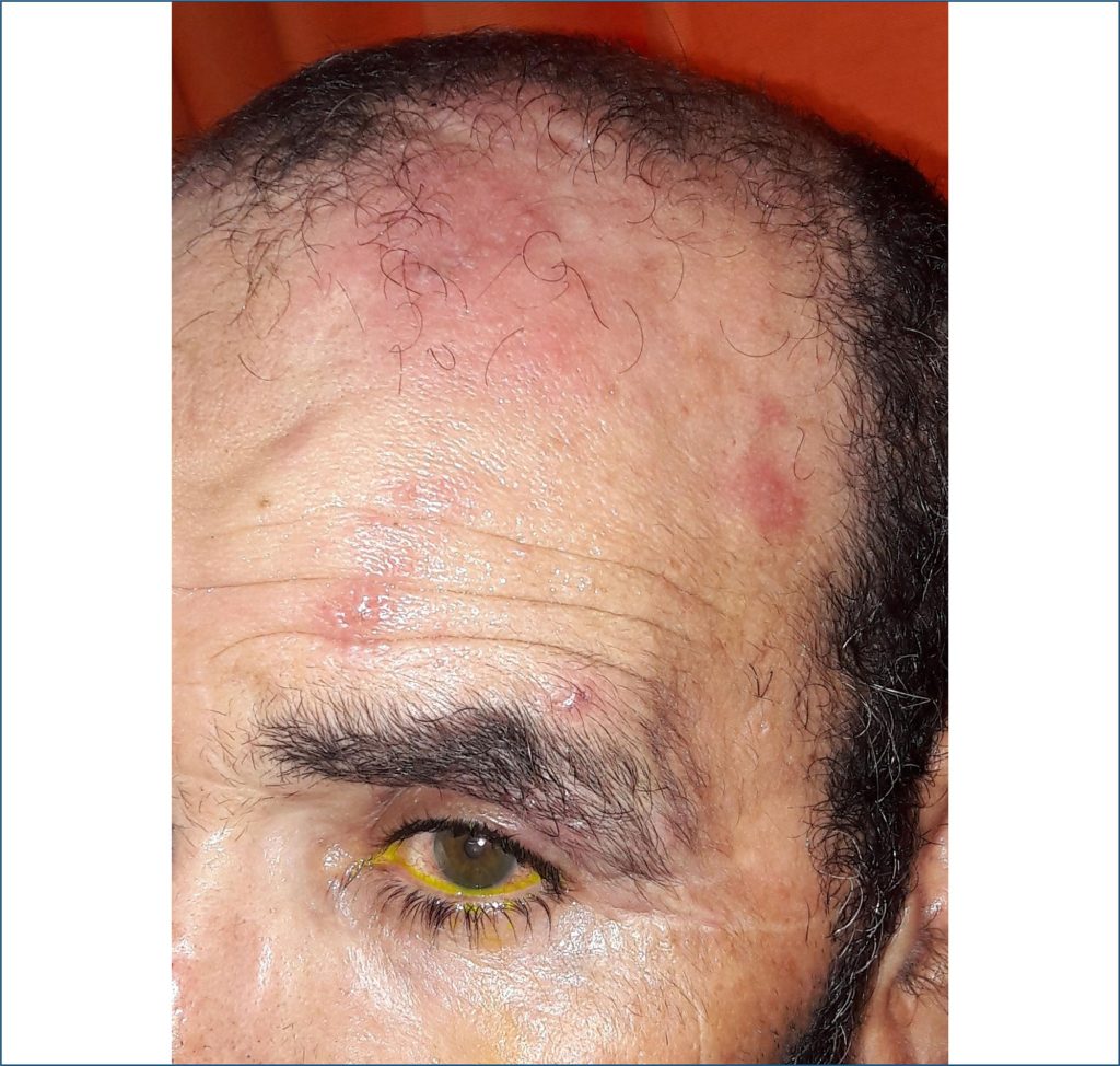
A 62-year-old healthy man with left-sided headaches for 3 days and redness of the left eye for 24 hours. The best corrected visual acuity of both eyes was 10/10. The left side of the forehead showed incipient vesicles on an erythematous background (). The left eye was slightly injected. Biomicroscopic examination disclosed a large temporal corneal pseudodendrite (). Fluorescein staining under a cobalt-blue filter confirmed this pattern (). Valacyclovir associated with lubricant eyedrops was started. Follow-up examination revealed complete resolution of symptoms and no corneal sequelae; the visual acuity was unaffected.
Herpes zoster ophthalmicus occurs when herpes zoster presents in the ophthalmic division of the fifth cranial nerve. Without the use of antiviral therapy, approximately 50% of herpes zoster patients develop ocular involvement.() Punctate and pseudo-dendritic types of keratitis are mainly observed during the early eruptive phase. Punctate epithelial keratitis corresponds to edematous epithelial cells where Herpes zoster replicates. Pseudodendrites are the results of the coalescence of previous punctate epithelial keratitis.() They are smaller and more superficially ulcerated than herpes simplex dendrites. They typically do not show terminal bulbs.
[…]
Keywords: Herpes Zoster Ophthalmicus; Pseudodendrites; Vesicles