Rev Bras Oftalmol.2025;84:e0082
Importance of confocal microscopy in the early diagnosis of fungal keratitis in contact lens users
DOI: 10.37039/1982.8551.20250082
ABSTRACT
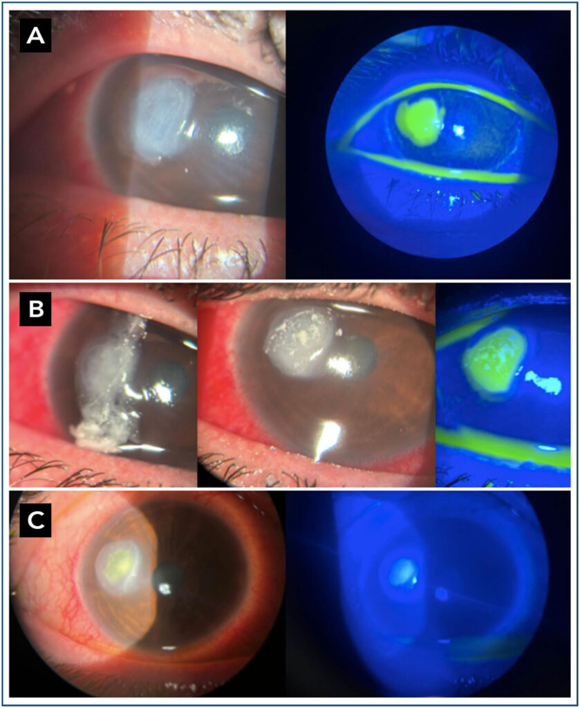
Fungal keratitis is a potentially sight-threatening corneal infection, often diagnosed late due to clinical overlap with other infectious etiologies. In vivo confocal microscopy has proven to be an effective method to detect fungal structures, even in the absence of microbiological confirmation. The objective was to report a case of fungal keratitis initially treated as bacterial, in which confocal microscopy was decisive for the etiological diagnosis and therapeutic reorientation. A 30-year-old female patient, regular contact lens wearer, presented with blurred vision, conjunctival hyperemia, and foreign body sensation in the right eye. Slit-lamp examination revealed a 2.2 mm peripheral corneal ulcer, which progressed to 3.4 mm after five days of empirical antibiotic therapy with no clinical improvement. Confocal microscopy showed filamentous structures suggestive of fungal infection, prompting the initiation of topical antifungal therapy (natamycin, later replaced by amphotericin B). Complete resolution of the lesion was achieved after 25 days of treatment. Confocal microscopy proved crucial for therapeutic reorientation, demonstrating its value in the early diagnosis of refractory keratitis.