We report a case of a 57-year-old woman with a 23-year history of systemic lupus erythematosus treated with hydroxychloroquine at a daily dose of 400 mg/d (6.51 mg/ kg/day), resulting in a cumulative dose of 3,358 g.
[…]
Multimodal imaging in a case of hydroxychloroquine induced maculopathy
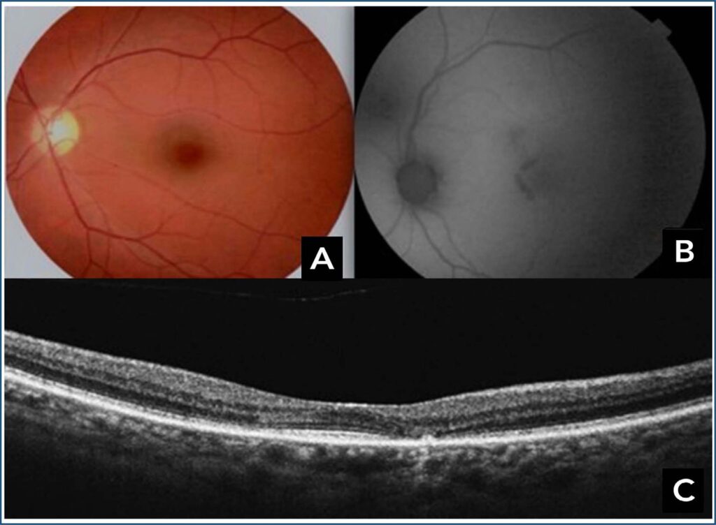
Figure 1
(A) Color fundus photograph showing a "bull's eye" maculopathy.
(B) Autofluorescence image showing a macular cockade image. (C) B-scan of macular optical coherence tomography showing thinning of the outer layers with interruption of the ellipsoid zone and alteration of the pigment epithelium in the parafoveolar area.
KhallouliA, SelmiS, OueslatiY. Multimodal imaging in a case of hydroxychloroquine induced maculopathy. Rev. bras.oftalmol. 2025;84:e0046.
Khallouli,Asma; Selmi,Slim; Oueslati,Yassin. Multimodal imaging in a case of hydroxychloroquine induced maculopathy. Rev. bras.oftalmol., v. 84, e0046, Jul. 2025.
Khallouli,A., Selmi,S., & Oueslati,Y. (2025). Multimodal imaging in a case of hydroxychloroquine induced maculopathy. Rev. bras.oftalmol.,84, e0046.
Khallouli,Asma and Selmi,Slim and Oueslati,Yassin. Multimodal imaging in a case of hydroxychloroquine induced maculopathy. Rev. bras.oftalmol. [online]. 2025, vol. 84, [cited 2026-03-08], e0046. Available from: <https://www.rbojournal.org/en/article/multimodal-imaging-in-a-case-of-hydroxychloroquine-induced-maculopathy/>. ISSN 0034-7280.