Rev Bras Oftalmol.2026;85:e0042
Optical coherence tomography characteristics of the optic nerve in Ecuadorian glaucoma patients
DOI: 10.37039/1982.8551.20260042
ABSTRACT
Objective:
To characterize structural optic nerve changes in Ecuadorian glaucoma patients using optical coherence tomography data from 2015 to 2022.
Methods:
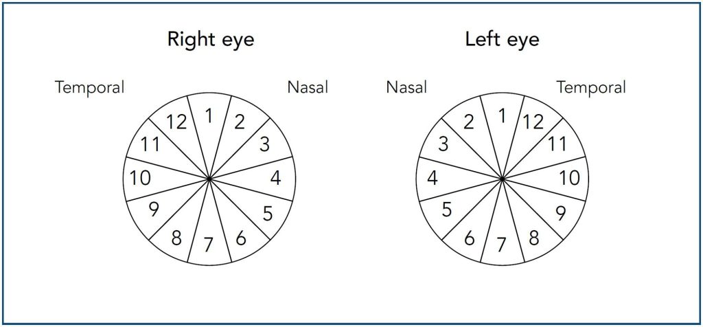
This retrospective study analyzed anonymized optical coherence tomography data from 301 eyes of 162 patients diagnosed with primary open-angle glaucoma. Data were acquired using the Nidek RS-330 and analyzed with Statistical Package for the Social Sciences, version 21. We assessed peripapillary retinal nerve fiber layer thickness (global, quadrants, and clock-hour sectors) and cup-to-disc ratios.
Results:
The mean global retinal nerve fiber layer thickness was 72.97 ± 16.48 μm (range 34 to 116 μm). The most affected retinal nerve fiber layer quadrants, in descending order of deterioration, were superior, inferior, nasal, and temporal. Pathological changes were most prevalent in clock-hour sectors 1, 5, 7, 11, and 12. Pathological horizontal cup-to-disc ratios were found in over 90% of patients.
Conclusion:
Ecuadorian glaucoma patients showed significantly reduced retinal nerve fiber layer thickness and high rates of pathological optical coherence tomography parameters, particularly in superior/inferior quadrants and horizontal cup-to-disc ratios, suggesting a distinct neuropathy pattern in this population. This data is crucial for future studies on a distinct glaucomatous neuropathy pattern within the Ecuadorian population.