Rev Bras Oftalmol.2023;82:e0032
Racemose hemangioma presenting as a vitreous hemorrhage: case report
DOI: 10.37039/1982.8551.20230032
ABSTRACT
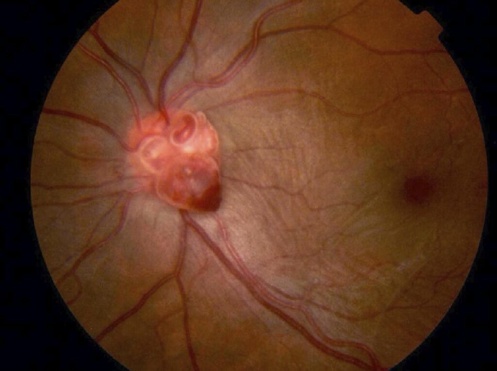
This report describes a case of retinal racemose hemangioma that first presented as a vitreous hemorrhage. The authors present the case of a 47-year-old woman with a sudden 5-day painless visual loss in her left eye. At the first visit, the best-correct visual acuities were 20/20 in the right eye and hand motions in the left eyes. Ultrasonography showed an attached retina and a massive vitreous hemorrhage. Pars plana vitrectomy was performed and a dilatation of large vessels was detected bulging from the optic disc. The best-correct visual acuities on day 30 postoperatively was 20/25 in the left eye. Fundus angiography and spectral-domain optical coherence tomography angiography showed anomalous arteriovenous communications with no intervening capillaries. The diagnosis was racemose hemangioma, an arteriovenous malformation of group 2 retina based on the Archer classification.