Rev Bras Oftalmol.2026;85:e0059
Evaluating peripheral retina: a cautionary note on miotics
DOI: 10.37039/1982.8551.20260059
ABSTRACT
Objective:
To underscore the critical importance of assessing the peripheral retina in all patients, especially those planning to use miotics.
Methods:
A retrospective and observational study was conducted at a single ophthalmological medical center, using records from the International Classification of Diseases (ICD-10) for closed-angle glaucoma and lesions predisposing to detachment. Participants in this study underwent anterior chamber assessment using the Goldman 3-Mirror lens and the Volk G-4 Gonioscopy lens. Peripheral retinal evaluation was conducted with indirect binocular ophthalmoscopy and posterior biomicroscopy, also utilizing the Goldman 3-Mirror lens. The medical records were analyzed, and all information was recorded, including procedures.
Results:
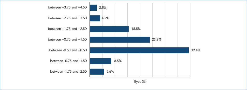
Among 9,854 patients assessed, 1,144 (11.6%) were classified with primary angle closure suspect, primary angle closure, or primary angle closure glaucoma. Additionally, 1,032 patients exhibited lesions predisposing to retinal detachment, constituting 10.4% of the cohort. Notably, 71 eyes from 56 (0.56%) patients demonstrated an association between primary angle closure suspect, primary angle closure, or primary angle closure glaucoma and these predisposing lesions. Within the primary angle closure suspect, primary angle closure, or primary angle closure glaucoma group, 56 (4.89%) presented with retinal lattice degeneration, holes, and/or tears. The majority were female, over 40 years old, hyperopic, with unilateral involvement and symptomatic. The average follow-up duration was 6.4 years.
Conclusion:
Peripheral degenerative lesions that may lead to retinal detachment are relatively common and can be found even in selected cases of angular narrowing. Peripheral retinal evaluation may be advisable in angle-closure eyes, particularly when miotics are planned; prospective studies are needed to confirm risk.