Rev Bras Oftalmol.2024;83:e0067
Confocal microscopy as an aid in the diagnosis of keratitis due to Candida parapsilosis
DOI: 10.37039/1982.8551.20240067
ABSTRACT
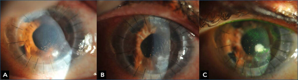
To describe a case of treatment-resistant corneal infiltrate in a patient 4 months after penetrating keratoplasty. The etiological diagnosis was established through microbiology and in vivo corneal confocal microscopy. A 80-year-old female patient presented with stromal infiltrate in the donor corneal bud 4 months following penetrating keratoplasty. Initially, the corneal culture was positive for Candida parapsilosis. In vivo corneal confocal microscopy demonstrated approximately 4 μm hyperreflective spherical microorganisms with a suggestive Candida spp. A second corneal culture was positive for Candida sp., and hyphae presence in the analyzed material was observed. The patient remained refractory to therapy. Confocal microscopy of the cornea continues to grow as a useful, non-invasive diagnostic method and in monitoring response to therapy. This report contributes by detecting Candida sp. in in vivo corneal confocal microscopy to aid in the diagnosis of keratitis.
Keywords: Candida parapsilosis; confocal; Corneal diseases; Fungi; keratitis; Keratoplasty; Microscopy; penetrating